Ultrasound & Elastography
What is ultrasound?
Ultrasound imaging uses sound waves to produce pictures of muscles, tendons, ligaments and joints throughout the body. It is used to help diagnose sprains, strains, tears, and other soft tissue conditions. Ultrasound is safe, noninvasive, and does not use ionizing radiation.
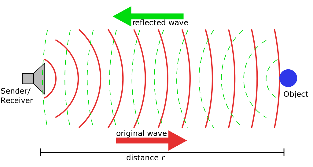
How does ultrasound work?
Ultrasound imaging involves the use of a transducer (probe) and ultrasound gel placed directly on the skin. High-frequency sound waves are transmitted from the probe through the gel into the body. The transducer collects the sounds that bounce back and a computer then uses those sound waves to create an image. Ultrasound examinations do not use ionizing radiation and there is no radiation exposure to the patient. Because ultrasound images are captured in real-time, they can show the structure and movement of the body.
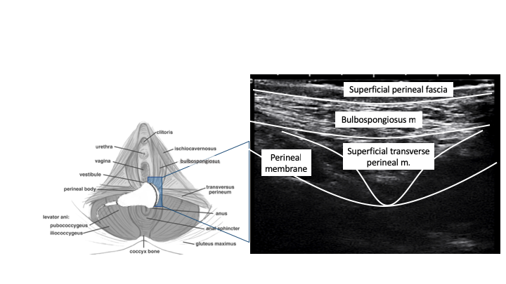
What can ultrasound tell us about fascia?
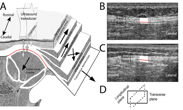
Real time ultrasound can be used to effectively rate the quality of connective tissue. The Cronbach’s alpha coefficient is 0.98, indicating that the use of ultrasound for rating of fascia has high inter-rater reliability. Organised fascia is defined as being able to draw a rectangular box around the hyperechoic zone of deep fascia. Disorganized is described as not being able to draw a rectangular box around the hyperechoic zone of deep fascia.
De Coninck K, Hambly K, Dickinson JW, Passfield L. Measuring the morphological characteristics of thoracolumbar fascia in ultrasound images: an inter-rater reliability study. BMC Musculoskelet Disord. 2018 Jun 1;19(1):180.
Langevin HM, Stevens-Tuttle D, Fox JR, Badger GJ, Bouffard NA, Krag MH, Wu J, Henry SM. Ultrasound evidence of altered lumbar connective tissue structure in human subjects with chronic low back pain. BMC Musculoskelet Disord. 2009;10:151.
What is ultrasound elastography?
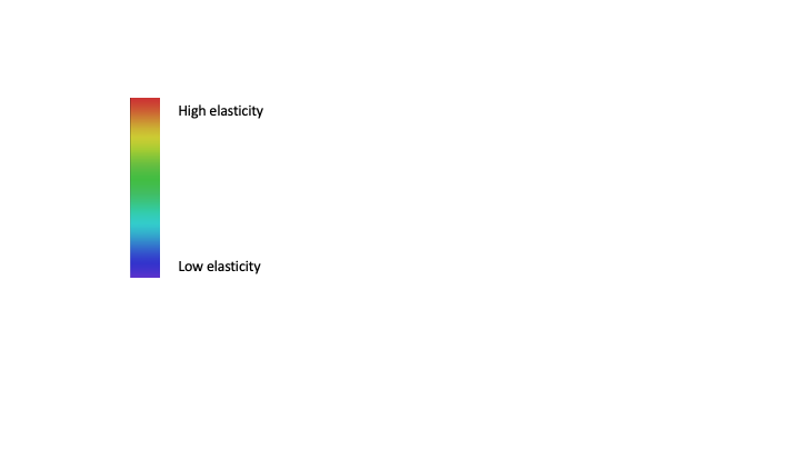
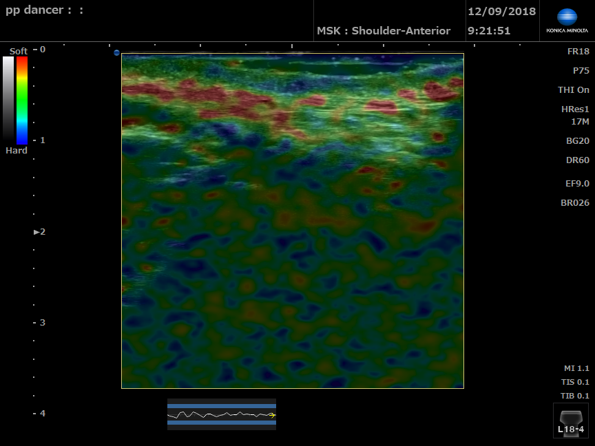
Ultrasound elastography is used to assess tissue stiffness. Strain images are generated by using the transducer to apply repetitive minimal pressure to the tissues. Under an equal amount of stress/tissue displacement, a stiff region experiences less deformation than surrounding softer tissue. A color map shows the different magnitudes of strain and relative stiffness of the tissue.
Small amplitude movements at strain of 0.1–2% in the tissue is sufficient. A real-time feedback indicator signifies whether the appropriate amount of stress is being applied.
The subsequent tissue displacement is tracked by computer software and the strain calculated from the axial gradient of the displacements.
This can aiding the assessment of the spatial relationship between the ultrasound image and the elastography data.